
Calcaneal Fat Pad Atrophy
Overview:
- Decreased elastic tissue
- Decreased water concentration
Anatomy:
Risk Factors:
- After approximately age 40, the fat pad begins to deteriorate
- Decreased elastic tissue
- Decreased water concentration
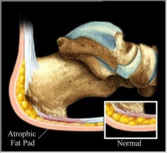
- The overall thickness and height of the fat pad decrease
- Each heel strike generates force equal to 110% of the body weight
- During running, heel strike can generate force equal to 250% of the body weight
Anatomy:
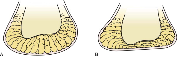
- Honeycombed pattern of fibroelastic septa
- These spaces are enclosed and contain fat globules
- The tissue septa form in a U-shape and attach to the calcaneus and skin
- Elastic transverse and diagonal fibers help reinforce the chambers internally
- The fat pad provides cushion to the hindfoot while keeping mechanical integrity for its shock absorption
Risk Factors:
- Increased age due to fat pad deterioration
- Obesity and subsequent increased pressure
- Genetics
- Steroid injections
- Prolonged standing or walking on hard surfaces with inappropriate footwear
- Excessive heel strike with poor footwear
|
Diagnosis/Classification:
|
|
Tests
Palpation of the fat pad
Observation
Diagnostic Imaging (Ultrasound or MRI)
Palpation of the fat pad
- softened and flattened surface
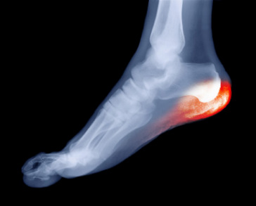
Observation
- erythema and inflammation over plantar aspect of the heel
Diagnostic Imaging (Ultrasound or MRI)
- Decreased thickness and height of fat pad
References:
- Prichasuk S. The heel pad in plantar heel pain. J Bone Joint Surg Br. Jan 1994;76(l):140-142
- Sawyer G, Lareau C, Mukand J. Diagnosis and Management of Heel and Plantar Foot Pain. Medicine & Health Rhode Island [serial online]. April 2012;95(4):125-128. Available from: Academic Search Complete, Ipswich, MA.
- DiGiovanni, Benedict F., Dawson, Laura K., Baumhauer, Judith F. Mann’s Surgery of the Foot and Ankle, Chapter 13, Pages 685-701
Last Edited by: Joseph Kauffman, SPT at AT Still University on July 12, 2014
